Acridine orange (AO) stained embryos (8-day mouse embryo) can be visualized to yield data that is similar to that obtained with SEM (Figure 13). AO was used to stain the embryos resulting in a surface fluorescence reflection. Embryos that are millimeters in thickness can be observed. The methodology has the advantage of reducing potential SEM artifacts of dehydration and critical point drying. Magnification 100x
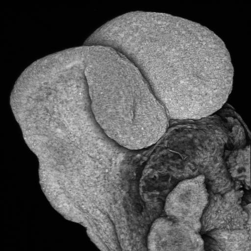 Figure 13
Circulatory system of 14th-day control rat limb (Figure 14) magnification 100x.
 Figure 14