Confocal Images of Reproduction
Robert M Zucker, Owen Price, Sid Hunter, John Rogers, Chris Lau and Jerry Goldman
Reproductive Toxicology Division, National Health and Environmental Effects Research Laboratory United States Environmental Protection Agency.
Ovaries
Ovaries were fixed with paraformaldehyde and stained with YO-PRO, dehydrated with MEOH, and cleared with BABB. The procedure allows for the visualization individual sections inside a follicle contained in an ovary. Magnification of the follicle is 100x while magnification of the oocyte is 600x. A 10 x objective was used to obtain the image of the follicle while a 20 x objective zoomed 3x was used to obtain the image of oocyte contained inside the follicles
 Figure 1 - description of oocyte contained in follicle
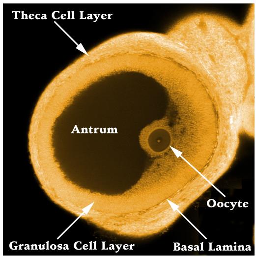 Figure 2 - description of individual follicle parts
 Figure 3 - nine middle sections of the follicle – note oocyte contained in the middle sections and enlarged in figures 1 and 2.
 Figure 4 - 25 sections of the follicle. Note the top and bottom and egg contained in the middle sections.