The tubules from a testis were stained with ethidium bromide. Figure 5 is an individual slice of a testis. Figure 6 shows the signal attenuation that occurs as one penetrates deep into tissues . In contrast Figure 7 is a seminiferous tubules that was stained with ethidium bromide. The corresponding sections of this structure, (Figure 8) shows less signal attenuation as the tissue is less dense. Magnification 100x
 Figure 5 Figure 5 |
 Figure 6 Figure 6 |
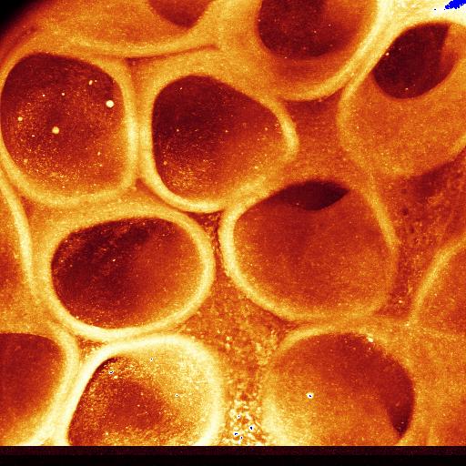 Figure 7 Figure 7 |
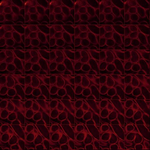 Figure 8 Figure 8 |