
Flow Cytometry and Microbiology
Rapid flow cytometric characterization of Escherichia coli.
Sergio Sgorbati
Sezione Botanica generale
Dipartimento di Biologia
Universita di Milano
Via Celoria 26
I-20133 Milano, Italy
 email: sgorbati@imiucca.csi.unimi.it
Silvia Barbesti
Bio-Rad Laboratories
Via Conca Del Naviglio 10
20123 Milano, Italy
email: Silvia_Barbesti@bio-rad.com
Data Examples
The following examples were collected using the Bio-Rad Bryte HS flow cytometer.
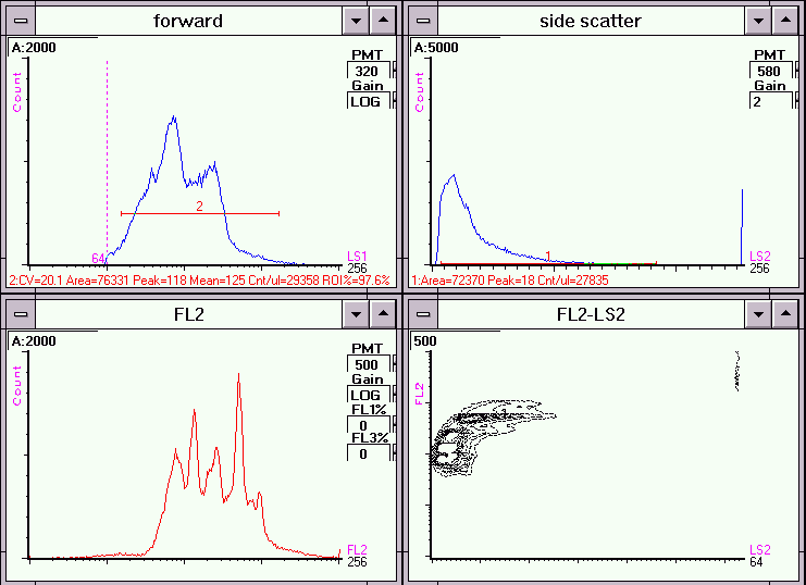
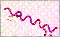
Figure 1. DNA staining of Escherichia coli cells using a combination of mithramycin and ethidium bromide.
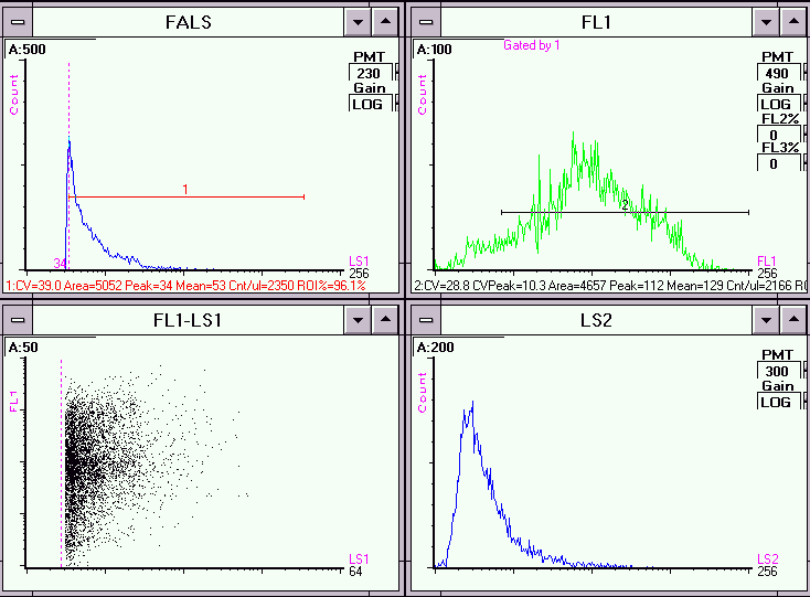
Figure 2a. Light scatter and fluorescence histograms for E.coli cells stained with the lipophilic cationic dye rhodamine 123.
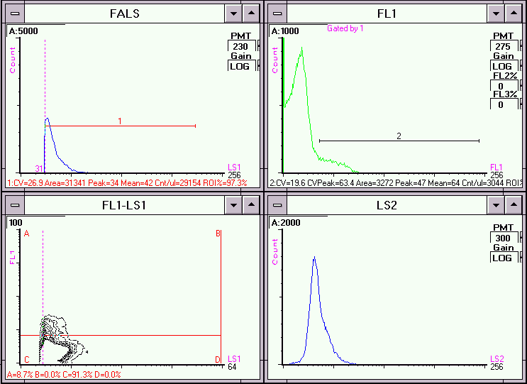
Figure 2b. Light scatter and fluorescence histograms for E.coli cells heat incubated at 80 degrees Celsius and stained with rhodamine 123.
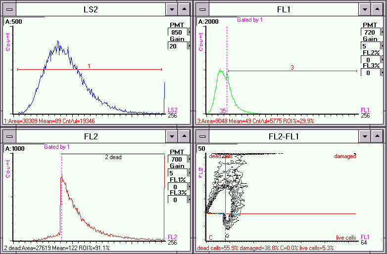
Figure 3. Light scatter and fluorescence histograms for E.coli cells heat incubated at 80 degrees Celsius and stained with DAPI and propidium iodide.
 Back to Flow Cytometry and Microbiology Introductory Page
CD ROM Volume 4 was produced by staff at the Purdue University Cytometry Laboratories
and distributed free of charge as an educational service to the cytometry community.
If you have any comments please direct them to
Dr. J. Paul Robinson, Professor & Director, PUCL, Purdue University, West Lafayette,
IN 47907. Phone:(765) 494-0757; FAX (765) 494-0517; Web http://www.cyto.purdue.edu EMAIL cdrominfo@flowcyt.cyto.purdue.edu