
Four color analysis on the B-D FACSCalibur.
Our Becton-Dickinson FACSCalibur has had all (most?) of the bugs worked out of it (including a new EPROM, for more consistent connections) and is now being routinely used by several laboratories for three- and four-color analysis of flow samples. As you may recall, this instrument differs from the FACScan in that it has two lasers: a conventional argon laser emitting at 488 nm, for excitation of FITC, PE, PI,etc. (as on the FACScan) and a diode laser emitting at 630 nm. This second laser makes possible simultaneous analysis of four fluorescent parameters (compared to the FAScan which can analyze only three). Thus, FITC, PE and PerCP-conjugated antibodies can be used in conjunction with a fourth red-emitting fluorochrome. Examples include allophycocyanin (APC) and Cy5, both of which can be excited by a diode laser and detected by the new instrument. As mentioned previously, the Flow Core has purchased limited quantities of APC- and Cy5-conjugated secondary antibodies and avidin to introduce users to the possibilities of 4-color phenotyping. We can also help you find commercial sources of APC- and Cy5-conjugated antibodies, and can help you conjugate Cy5 to your own antibodies and proteins. One laboratory (Dr. Lacy’s at Sloan-Kettering) is currently using the instrument to detect CD3, CD8, b220 and surface Ig simultaneously on transgenic mouse splenocytes (using APC, FITC, PE and PerCP-conjugated antibodies). Contact us for assistance in working out your own protocols.
(Below). The FACSCalibur with the "hood up". Argon and diode lasers are located in the black and yellow housing on the left. The flow cell and PMTs are located almost directly above the sample port. The mechanical sort module is the plexiglass array on the right. Access to the interior of the instrument (unlike the FACScan) allows us to modify the filter configuration of the instrument, if desired.

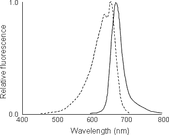
Allophycocyanin (APC) is a zooplankton-derived fluorochrome that can be coupled to a wide variety of proteins and antibodies. It is excited at 630 nm and has an emission maxima of approximately 660 nm. It is among the brightest fluorochromes per unit molecule known (exceeding even PE) and is very useful for surface marker immunophenotyping. It can be easily combined with FITC, PE and PerCP or RED 613 for four-color surface immunophenotyping on lymphocytes and other cell types. Even for two- and three-color analysis, the greater fluorescence of APC and the lower autofluorescence background generated by the diode laser make APC a good choice for the detection of weakly expressed surface markers.
(Below). Excitation (dotted line) and emission (solid line) spectra of APC.
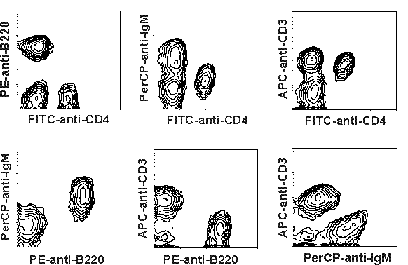
(Below). An example of a four-color experiment using FITC-anti-CD8, PE-anti-b220, PerCP-anti-IgM and APC-anti-CD3 to phenotype mouse splenocytes (courtesy of Tonya Villafana and Dr. Elizabeth Lacy). PE-Texas Red conjugates (including RED613 from Gibco) can be used in place of PerCP as the FL3 flurochrome. In some cases PE-Cy5 conjugates (sold under the names CyChrome, TriColor, etc.) can also be used for FL3; the Cy5 portion of the tandem conjugate is excited by the diode laser, however, and can interfere with the APC signal.
Cy5 is a cyanin dye that can be easily coupled to most proteins. Although dimmer than APC, it is more useful for applications where low molecular weight is preferred, such as staining for intracellular antigens. The coupling procedure for Cy5 is much easier than for APC and can be done using a commercial kit from Amersham (they have more information on Cy5 and other Cy series fluorochromes, too).
(Below). Excitation (dotted line) and emission (solid line) spectra of Cy5.

(Below). A comparison of Cy5 and FITC for the detection of cyclin A and B1 in HL-60 cells. Cy5 shows increased sensitivity for the detection of human cyclin proteins compared with FITC. Although Cy5 and FITC have comparable fluorescent intensities, lower cellular autofluorescence generated by the diode laser increases signal-to-noise ratio and overall sensitivity.

Back to the HSS Flow Cytometry Core Facility Home Page.