
The effects of microgravity and space flight on chick chondrocyte proliferation and differentiation.
(A collaboration with Dr. Stephen Doty at the Hospital for Special Surgery).
The Flow Core is using flow cytometric analysis of chick chondrocyte DNA content and cell cycle protein expression to supplement Dr. Doty's on-going studies into the effects of microgravity on chondrocyte proliferation and development. Chick chondrocytes can be mechanically disaggregated and the resulting single cell suspensions stained with a DNA binding dye such as propidium iodide. The resulting cells can be analyzed by flow cytometry to measure the percentage of cells in the G0 (quiescent), G1 (non-cycling but replication-competent), S-phase (replicating their DNA) and G2 and M (pre-mitotic and mitotic) phases. In addition, these cells can be immunophenotyped with fluorochrome-conjugated antibodies against important cell cycle regulatory proteins to provide further information about the status of the chondrocyte cell cycle. We have currently developed labeling methods for the immunodetection of cyclin D2 and D3 (a G1 and S phase regulatory protein), cyclin E (a G1 phase protein) and proliferating cell nuclear antigen (a component of the DNA polymerase delta complex). Dr. Doty is also developing the immunocytochemical techniques for detecting these proteins in sections of intact chondrocyte colonies. Simultaneous detection of DNA cell cycle and cyclin expression by flow cytometry and analysis of cyclin expression by immunocytochemistry will provide powerful and complementary tools for detecting subtle changes in the chondrocyte cell cycle induced by microgravity and space flight.
(Below). Flow histograms and cytograms of propidium iodide-labeled DNA content (top panels) and cyclin D2/D3 expression (lower panels) in chick chondrocytes (right panels) harvested at Day 10 following initial seeding of cultures. HL-60 human myeloid leukemia cells (left column), which also express cyclin D2 and/or D3, were run simultaneously as a control. Cyclins D2 and D3 are expressed late in G1 and throughout S phase, as illustrated here by their strong presence throughout the cell cycle. A portion of the cells included in Dr. Doty's October 1998 shuttle launch experiment will be analyzed in this fashion, supplementing morphological and immunocytochemical analysis. We will also be analyzing alkaline phosphatase in these cells using a flow-based assay.
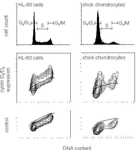
These samples were stained with anti-cyclin D antibody followed by a Cy5-conjugated secondary and analyzed on the FACSCalibur. The argon-diode dual laser allows simultaneous analysis of propidium iodide and Cy5, even though their emission spectra overlap. Cy5 appears brighter than FITC due to lower background autofluorescence when using a red laser, making it more sensitive than FITC for detection of the very low levels of cyclins found in chick chondrocytes. For more information on the FACSCalibur and diode laser-excited probes, click here.
![]() The
page is still under construction. Please check back soon for more
stuff!
The
page is still under construction. Please check back soon for more
stuff!
Back to the HSS Flow Cytometry Core Facility Home Page.